Spinal Cord Tracts
Three layers of meninges surround the spinal cord and spinal nerve roots. Central cord syndrome CCS Brown-Sequard syndrome BSS anterior cord syndrome ACS conus medullaris syndrome CMS cauda equina syndrome CES and posterior cord syndrome PCS.
1
Lab 2 Spinal Pathways

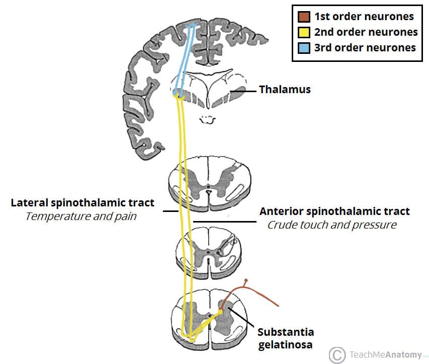
The Ascending Tracts Dcml Anterolateral Teachmeanatomy
Spinal Cord Nuclei.

Spinal cord tracts. Dura mater consists of two layers-. Subacute combined degeneration of the spinal cord is a neurological complication of vitamin B12 cobalamin deficiency. The tip of the spinal cord is called the conus.
The spinal nerves below the level of injury get signals but they are not able to go up the spinal tracts to the brain. Subacute combined degeneration is characterized by degeneration of the dorsal columns. The spinal cord is a tubular bundle of nervous tissue and supporting cells that extends from the brainstem to the lumbar vertebraeTogether the spinal cord and the brain form the central nervous system.
Commissural tracts enable the left and right sides of. We have 31 pairs of spinal nerves. Spinal nerves act as mediators communicating information to and from the rest of the body and the spinal cord.
In human the spinal cord ends at L2 vertebral level. Intramedullary cord hyperintensity at T2-weighted MRI is a common imaging feature of disease in the spinal cord but it is nonspecific. These are bundled into specialized tracts that conduct impulses triggered by pressure pain heat and other sensory stimuli or conduct motor impulses activating muscles and glands.
Commissural tracts cross from one cerebral hemisphere to the other through bridges called commissures. In each of the spinal cords many segments lives a pair of roots that are made up of nerve fibers. The spinal cord is a long thin tubular structure made up of nervous tissue which extends from the medulla oblongata in the brainstem to the lumbar region of the vertebral columnIt encloses the central canal of the spinal cord which contains cerebrospinal fluidThe brain and spinal cord together make up the central nervous system CNS.
Symptoms may include loss of muscle function sensation or autonomic function in the parts of the body served by the spinal cord below the level of the injury. Marginal zone MZ posterior marginalis located at the tip of the dorsal horn and is important for relaying pain and temperature sensation to the brain. The prominent nuclei groups of neuron cell bodies in the spinal cord are the.
The spinal cord is more than just a conduit as it also modifies and. The spinal cord has a core of tissue containing nerve cells surrounded by long tracts of nerve fibers consisting of axons. The spinal cord is part of the central nervous system CNS which extends caudally and is protected by the bony structures of the vertebral column.
While central cord syndrome is the most common SCI syndrome the less commonly. It is an emergency which can require urgent surgical intervention to prevent long-term. Tracts in your spinal cord carry messages between your brain and the rest of your body.
It conveys proprioceptive information and on-going activity in the spinal cord interneurons. A spinal cord injury SCI is damage to the spinal cord that causes temporary or permanent changes in its function. Spinocerebellar Tract - From spinal cord interneurons.
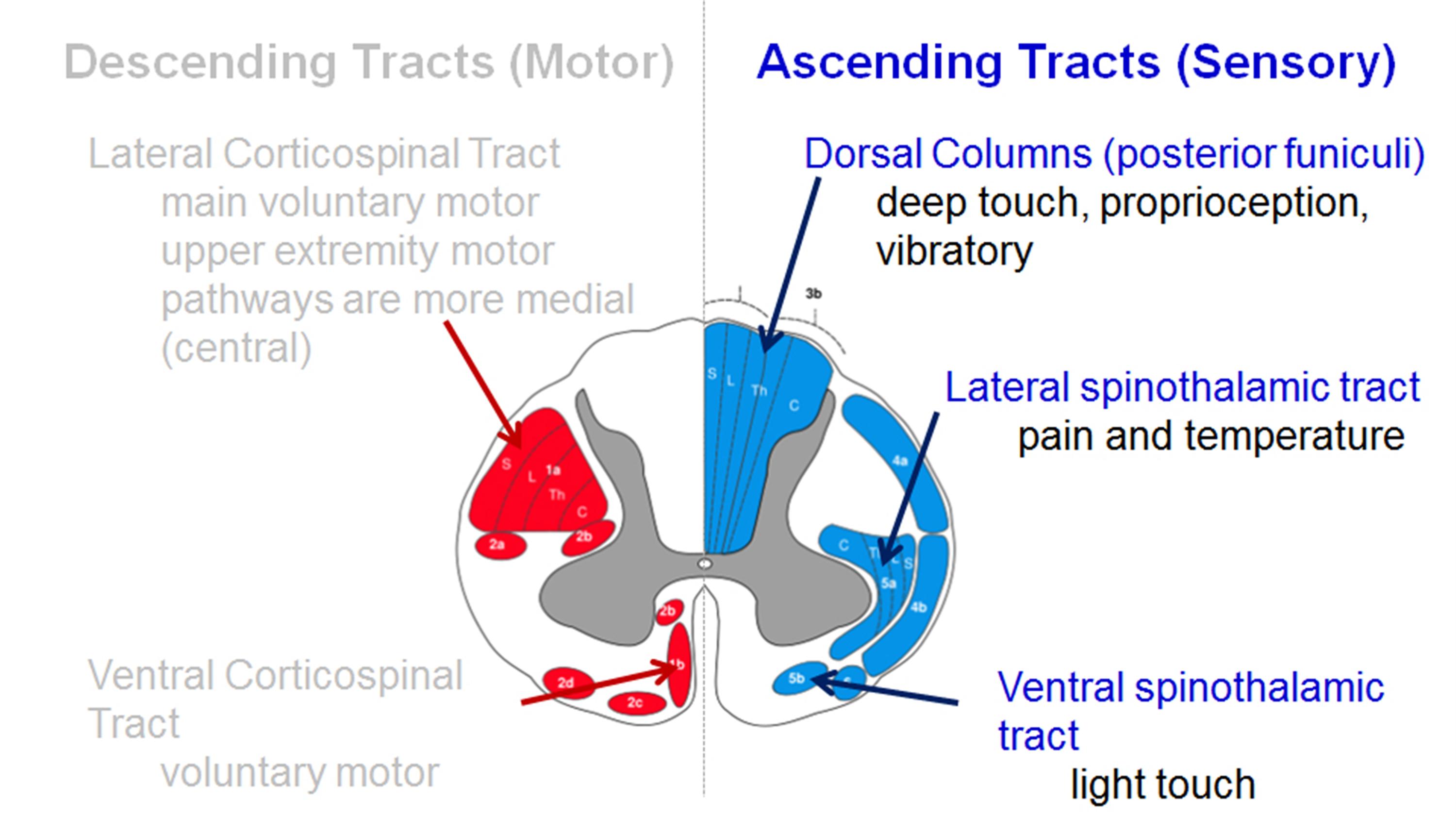
Injury can occur at any level of the spinal cord and can be complete with a total loss of sensation and muscle. Spinal Cord Cresyl Violet Cross section of the spinal cord stained with cresyl violet. Spinal cord tracts diagram Ascending tracts convey information from the periphery to the brain.
The spinal cord is a complex cylinder of nerves that starts at the base of your brain and runs down the vertebral canal to the backbone. Radiologists play a valuable role in helping narrow the differential diagnosis by integrating patient history and laboratory test results with key imaging characteristics. Spinothalamic tracts cross at spinal cord level classically 2-levels below Prognosis.
Motor tracts carry signals from your brain to control muscle movement. When the spinal cord is damaged the message from the brain cannot get through. Traumatic spinal cord injury can manifest as a wide variety of clinical syndromes resulting from damage to the spinal cord or its surrounding structures.
99 ambulatory at final follow up. It can result from minor injury if the spine is weakened from disease such as ankylosing spondylitis or if there is pre-existing spinal stenosis. The lumbosacral spinal cord however starts at about T9 and continues only to L2.
In this article we shall examine the macroscopic anatomy of the spinal cord its structure membranous coverings and blood supply. Below this region is a group of nerve roots called the cauda equina. The spinal cord is the major conduit through which motor and sensory information travel between brain and body.
The outer layer of the human spinal cord consists of white matter ie myelin-sheathed nerve fibers. The corticospinal tracts for example carry motor signals from the cerebrum to the brainstem and spinal cord. It is covered by the three membranes of the CNS ie the dura mater arachnoid and the innermost pia mater.
In cross-section the spinal cord is divided into the butterfly-shaped grey matter and surrounding white matter. It contains most of the segments that innervate the hip and legs as well as the buttocks and anal regions. In humans the spinal cord begins at the occipital.
Then a subset of SCIs is classified by their clinical presentation into 6 Spinal Cord Injury Syndromes. The spinal cord has numerous groups of nerve fibers going towards and coming from the brain. It has two tracts a Posterior Spinocerebellar Tract which relays via inferior cerebellar peduncle and b Anterior Spinocerebellar Tract relays via superior cerebellar peduncle to the cerebellum.
The tracts extend up and down the spinal. The tracts are responsible for carrying sensory and motor stimuli to. These have been collectively called the ascending and descending tracts of the spinal cord respectively.
The authors present an algorithmic approach to evaluating intrinsic abnormality of. The spinal cord provides a two-way conduction pathway to and from the brain and it is a major reflex center spinal reflexes are completed at this level. Enclosed within the vertebral column the spinal cord extends from the foramen magnum of the skull to the first or second lumbar vertebra where it ends just below the ribs.
A deficiency of vitamin B12 can occur as a result of nutritional deficiency reduced absorption due to altered gastrointestinal anatomy or function or due to the intake of certain drugs. It is part of the bodys collection of nerves called the central nervous system along with the brain. Motor pain light touch.
Best prognosis for function motor activity. The spinal cord contains longitudinally oriented spinal tracts white matter surrounding central areas gray matter where most spinal neuronal cell bodies are located. Substantia gelatinosa SG located at the top of the dorsal horn the SG is important for relaying pain temperature and light touch.
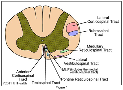
Normally messages are sent from the brain through the spinal cord to parts of the body which leads to movement. On the other hand the descending tracts carry information from the brain to the periphery. Figure 155 The Corticospinal Tracts and Other Descending Motor Tracts in the Spinal Cord KEY Axon of upper- motor neuron Lower-motor neuron Motor homunculus on primary motor cortex of left cerebral hemisphere Corticobulbar tract Cerebral peduncle MESENCEPHALON MEDULLA OBLONGATA Pyramids Decussation of pyramids To.
In most adult mammals it occupies only the upper two-thirds of the vertebral canal as the growth of the bones composing the vertebral. Cresyl violet is a basic dye that binds nucleic acids and preferentially stains RNA. The lower end of your spinal cord stops a little above your waist in the region called the conus medullaris.
Spinal Cord Anatomy Spine Orthobullets
Major Ascending And Descending Tracts In The Spinal Cord Sciencedirect

Spinal Cord Tracts And Reflexes Knowledge Amboss
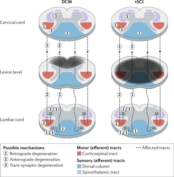
Traumatic And Nontraumatic Spinal Cord Injury Pathological Insights From Neuroimaging Nature Reviews Neurology

Sensory And Motor Spinal Tracts Of The Human Spinal Cord

Spinal Cord Tracts 600x274 Png Download Pngkit
Neuroanatomy Lab Online Lab 9 Page 32
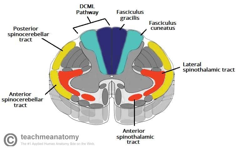
The Ascending Tracts Dcml Anterolateral Teachmeanatomy
0 Response to "Spinal Cord Tracts"
Post a Comment